The fornix (meaning "arch" in Latin) is a C-shaped bundle of nerve fibers in the brain that acts as the major output tract of the hippocampus. The fornix also carries some afferent fibers to the hippocampus from structures in the diencephalon and basal forebrain. The fornix is part of the limbic system. While its exact function and importance in the physiology of the brain are still not entirely clear, it has been demonstrated in humans that surgical transection – the cutting of the fornix along its body – can cause memory loss. There is some debate over what type of memory is affected by this damage, but it has been found to most closely correlate with recall memory rather than recognition memory. This means that damage to the fornix can cause difficulty in recalling long-term information such as details of past events, but it has little effect on the ability to recognize objects or familiar situations.
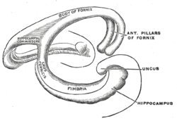

Structure
Functional Consequences of Fornix Damage
The fornix is essential for acquiring and consolidating new episodic memories. Fornix transection studies in macaques[1] have shown that the monkeys were strongly impaired on object-in-scene learning, which is a type of recall memory, specifically episodic-like memory (integrating what+where, although not when). Fornix transection in rodents impairs performance on tasks that require the encoding and retrieval of spatiotemporal context and, therefore, serves as a proxy for human episodic memory. For instance, fornix transection consistently leads to robust impairments in learning new routes and spatial locations (reviewed by [2]).
Fornix damage in humans is rare; a few individuals have had their fornix transected inadvertently during removal of colloid cysts from their third ventricles.[3] Nevertheless this small literature has consistently reported a persistent anterograde amnesia that is indistinguishable from the anterograde amnesia observed after focal hippocampal lesions. Deficits in recall are greater than for recognition, and the deficit is found across all types of material (e.g. visual and verbal) (reviewed by [2]). This supports the idea that damage to any part of the extended hippocampal memory system causes similar memory deficits.[4] Other aspects of cognition, such as social cognition and language ability, remain intact after fornix damage.
Lesion findings have been extended by work using the non-invasive in vivo technique diffusion-weighted imaging. This literature has shown that fractional anisotropy (FA) in the fornix decreases with advanced age, correlates with age-related memory impairments, and is relatively decreased in mild cognitive impairment and in Alzheimer's Disease (reviewed by [5][2]). New research studies are using deep brain stimulation to stimulate the fornix as some evidence has shown that doing so improves episodic memory (reviewed by [6]
Function
The fornix is the conduit by which the neurotransmitter acetylcholine –which is crucial for memory encoding–is sent from the medial septum/Diagonal Band of Broca to the hippocampus (reviewed in [7]). In addition, the GABA-producing neurons in the septal nuclei generate theta rhythms which are transmitted through the fornix to the hippocampus.[8][9] In the absence of these external modulators, the hippocampus is radically dysfunctional. In addition, the fornix transmits mnemonic information from the hippocampus to deep brain structures, which potentially allows us to use stored memories to guide us to rewarding people, places, and sources of sustenance.
Additional images
| This article uses material from the Wikipedia article Metasyntactic variable, which is released under the Creative Commons Attribution-ShareAlike 3.0 Unported License. |