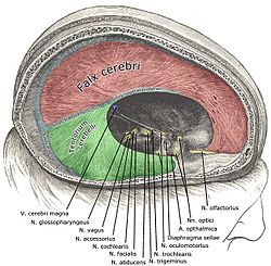
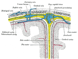
The falx cerebri, also known as the cerebral falx, is a large, crescent-shaped fold of meningeal layer of dura mater that descends vertically in the longitudinal fissure between the cerebral hemispheres of the human brain. The falx cerebri attaches anteriorly at the crista galli in proximity to the cribriform plate and to the frontal and ethmoid sinuses. Posteriorly, it is connected with the upper surface of the cerebellar tentorium. Its superior margin is attached at midline to the internal surface of skull, as far back as the internal occipital protuberance. The superior sagittal sinus is contained in the superior margin of the falx cerebri and overlies the longitudinal fissure of the brain. The inferior sagittal sinus is contained in the inferior margin of the falx cerebri and arches over the corpus callosum, deep in the longitudinal fissure.[1] The falx cerbri is named for its sickle-like form.
Calcification
Calcification of the falx cerebri is more prevalent in older patients, often without a determinable cause, and without pathogenic symptoms.[2]
Meningioma
Falcine meningioma is a meningioma arising from the falx cerebri and completely concealed by the overlying cortex. Falcine meningioma tends to grow predominately into one cerebral hemisphere but is often bilateral, and in some patients the tumor grows into the inferior edge of the sagittal sinus. However, although much information is available regarding meningiomas, little is known about falcine meningiomas.[3]
Additional images
This article uses material from the Wikipedia article
Metasyntactic variable, which is released under the
Creative Commons
Attribution-ShareAlike 3.0 Unported License.
Metasyntactic variable, which is released under the
Creative Commons
Attribution-ShareAlike 3.0 Unported License.